Quantcast
Channel: Caltech News tagged with "biology"

Learning from Mistakes

$
0
0
Discovering the neurons that rapidly catch our mistakes and how they correct our behavior
News Writer: 
Lori Dajose

RAdolphs Neurons Monitor for Mistakes FNL
RAdolphs Neurons Monitor for Mistakes FNL
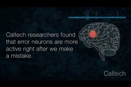
Researchers in the Adolphs laboratory at Caltech have discovered that certain types of neurons called error neurons are more active when we make a mistake. Take the Stroop test and see how you fare.
Credit: Caltech

Everyone makes little everyday mistakes out of habit—a waiter says, "Enjoy your meal," and you respond with, "You, too!" before realizing that the person is not, in fact, going to be enjoying your meal. Luckily, there are parts of our brains that monitor our behavior, catching errors and correcting them quickly. 

A Caltech-led team of researchers has now identified the individual neurons that may underlie this ability. The work provides rare recordings of individual neurons located deep within the human brain and has implications for psychiatric diseases like obsessive-compulsive disorder.

The work was a collaboration between the laboratories of Ralph Adolphs (PhD '93), Bren Professor of Psychology, Neuroscience, and Biology, and the Allen V. C. Davis and Lenabelle Davis Leadership Chair and director of the Caltech Brain Imaging Center of the Tianqiao and Chrissy Chen Institute for Neuroscience; and Ueli Rutishauser (PhD '08), associate professor of neurosurgery, neurology, and biomedical sciences, and Board of Governors Chair in Neurosciences at Cedars-Sinai Medical Center. A paper describing the research was published online in the journal Neuron on December 4.

"Many people know the feeling of making a mistake and quickly catching oneself—for example, when you are typing and press the wrong key, you can realize you made a mistake without even needing to see the error on the screen," says Rutishauser, who is also a visiting associate in Caltech's Division of Biology and Biological Engineering. "This is an example of how we self-monitor our own split-second mistakes. Now, with this research, we know which neurons are involved in this, and we are starting to learn more about how the activity of these neurons helps us change our behavior to correct errors."

In this work, led by Caltech graduate student Zhongzheng (Brooks) Fu, the researchers aimed to get a precise picture of what happens on the level of individual neurons when a person catches themselves after making an error. To do this, they studied people who have had thin electrodes temporarily implanted into their brains (originally to help localize epileptic seizures). The work was done in collaboration with neurosurgeon Adam Mamelak, professor of neurosurgery at Cedars-Sinai, who has conducted such electrode implantations for clinical monitoring of epilepsy for over a decade and closely collaborated on the research studies.

While neural activity was measured in their medial frontal cortex (MFC), a brain region known to be involved in error monitoring, the epilepsy patients were given a so-called Stroop task to complete. In this task, a word is displayed on a computer screen, and the patients are asked to identify the color of the text. Sometimes, the text and the color are the same (the word "green" for example, is shown in green). In other cases, the word and the color are different ("green" is shown in red text). In the latter case, the correct answer would be "red," but many people make the error of saying "green." These are the errors the researchers studied.

The measurements allowed the team to identify specific neurons in the MFC, called self-monitoring error neurons, that would fire immediately after a person made an error, well before they were given feedback about their answer.

For decades, scientists have studied how people self-detect errors using electrodes placed on the surface of the skull that measure the aggregate electrical activity of thousands of neurons. These so-called electroencephalograms reveal that one particular brainwave signature, called the error-related negativity (ERN), is commonly seen on the skull over the MFC right after a person makes an error. In their experiments, Fu and his colleagues simultaneously measured the ERN as well as the firing of individual error neurons.

They discovered two fundamental new aspects of the ERN. First, an error neuron's activity level was positively correlated with the amplitude of the ERN: the larger the ERN for a particular error, the more active were the error neurons. This finding reveals that an observation of the ERN—a noninvasive measurement—provides information about the level of activity of error neurons found deep within the brain. Second, they found that this ERN–single-neuron correlation, in turn, predicted whether the person would change their behavior—that is, if they would slow down and focus more to avoid making an error on their next answer. If the error neurons fired but the brain-wide ERN signature was not seen or was weak, the person might still recognize that they made an error, but they would not modify their behavior for the next task. This suggests that the error neurons need to communicate their error detection to a large brain network in order to influence behavior. 

The researchers found further specific evidence for parts of the circuit involved.

"We found error neurons in two different parts of the MFC: the dorsal anterior cingulate cortex (dACC) and the pre-supplementary motor area (pre-SMA)," says Fu. "The error signal appeared in the pre-SMA 50 milliseconds earlier than in the dACC. But only in the dACC was the correlation between the ERN and error neurons predictive of whether a person would modify their behavior. This reveals a hierarchy of processing—an organizational structure of the circuit at the single-neuron level that is important for executive control of behavior."

The research could also have implications for understanding obsessive-compulsive disorder, a condition in which a person continuously attempts to correct perceived "errors." For example, some individuals with this condition will feel a need to repeatedly check, in a short time period, if they have locked their door. Some people with obsessive-compulsive disorder have been shown to have an abnormally large ERN potential, indicating that their error-monitoring circuitry is overactive. The discovery of error neurons might facilitate new treatments to suppress this overactivity. 

The researchers next hope to identify how the information from error neurons flows through the brain in order to produce behavioral changes like slowing down and focusing. "So far, we have identified two brain regions in the frontal cortex that appear to be part of a sequence of processing steps, but, of course, the entire circuit is going to be much more complex than that," says Adolphs. "One important future avenue will be to combine studies that have very fine resolution, such as this one, with studies using fMRI [functional magnetic resonance imaging] that give us a whole-brain field of view."

The paper is titled, "Single-neuron correlates of error monitoring and post-error adjustments in human medial frontal cortex." In addition to Fu, Adolphs, Rutishauser, and Mamelak, other co-authors are Caltech scientist Daw-An Wu; Ian Ross of the Huntington Memorial Hospital in Pasadena; and Jeffrey Chung of Cedars-Sinai Medical Center. Funding was provided by the National Institutes of Health, the National Science Foundation, and the McKnight Endowment Fund for Neuroscience. This study was approved by the institutional review boards of Caltech, Cedars-Sinai Medical Center, and Huntington Memorial Hospital.

Neuroscience Initiative Turns Two

$
0
0
Spearheading interdisciplinary brain research
News Writer: 
Lori Dajose
A rendering of the Chen Neuroscience Research Building.
Credit: Smith Group

Two years ago, the Tianqiao and Chrissy Chen Institute partnered with Caltech to launch a major neuroscience initiative. Central to the initiative was the creation of the Tianqiao and Chrissy Chen Institute for Neuroscience at Caltech, where research investigations span a continuum, from deciphering the basic biology of the brain to understanding sensation, perception, cognition, and human behavior, with the goal of making transformational advances that will inform new scientific tools and medical treatments.

Construction on the Chen Neuroscience Research Building began in March 2018 and is expected to be completed by October 2020.

Fast facts about the Chen Building:

  • Construction of the building had to be carefully scheduled around lamprey breeding season. The laboratory of Marianne Bronner [Albert Billings Ruddock Professor of Biology] uses the jawless fish for studying developmental biology, and any vibrations caused by construction could have disrupted their summer breeding.
  • To accommodate the building's basement and sub-basement, 67,000 cubic yards of dirt were excavated from the site.
  • The building will house labs and offices for more than a dozen principal investigators.
  • The floors in the Chen building are aligned with the floors in the Broad building to the south, facilitating a subterranean tunnel connection between the two buildings.
  • Chen construction required the removal of several existing facilities, including the Wilson Court bungalows, originally located at the southeast corner of Wilson Avenue and Del Mar Boulevard. In recognition of Pasadena's architectural heritage, Caltech elected to preserve the 1920s-era bungalows—and their historically significant configuration—rather than demolish them. The seven bungalows have been moved to the northeast corner of Catalina Avenue and San Pasqual Street and will serve as campus housing once renovations are complete in May of 2019. 

For more neuroscience stories, visit: http://www.caltech.edu/news/tag_ids/155

Regulating the Rapidly Developing Fruit Fly

$
0
0
A clever program modulates some of the rapid phases of Drosophila development
News Writer: 
Lori Dajose
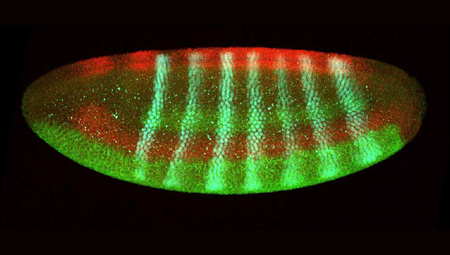
A Drosophila embryo at the point of cellularization, when it changes from a single "bag" of nuclei into individual cells.
Credit: Courtesy of the Stathopoulos laboratory

From birth, it takes humans almost two decades to reach adulthood; for a fruit fly, it takes only about 10 days. During a fly embryo's initial stages of development, the insect looks different from minute to minute, and its body plan is defined in just a few hours. Caltech researchers have now gained new insights into how a fly's genes influence this fast period of development—work that ultimately could shed light on the rapid cellular proliferation that occurs in other situations, including human cancers.  

The research was done in the laboratory of Professor of Biology Angelike Stathopoulos. A paper describing the research appears in the December 17 issue of the journal Developmental Cell.

Unlike a human embryo, which begins as a single cell that divides into more cells, the early embryo of the fruit fly Drosophila melanogaster is a single football-shaped bag of dividing nuclei. Each division of the nuclei constitutes one nuclear cycle, which takes between 8 and 15 minutes to complete. After the 14th nuclear cycle, the bag contains about 6,000 nuclei. At this point, the embryo separates into individual cells and the length of the cycles increases.

A nucleus contains an organism's genetic information, and can be thought of as a library. Each gene is like an individual reference book, containing instructions on how to build a protein. These books—the genes—never leave the library, so in order for the cell to use the instructions to build a protein, a photocopy must be made and taken out of the nucleus. This process of photocopying is called transcription.

There are limits to how quickly a gene can be transcribed, and so some especially long genes cannot be transcribed in their entirety during the limited amount of time that constitutes a single nuclear cycle. Instead, only a portion of the gene gets transcribed—only a chapter of the book is photocopied. 

For the Developmental Cell paper, a team of Caltech scientists led by former graduate student Jeremy Sandler (PhD '17) aimed to study what function, if any, these shorter transcripts have in the growing embryo.

In particular, the group studied the activity of one long gene with the counterintuitive name short gastrulation, or sog, during the early nuclear cycles of Drosophila embryos. The sog gene encodes a complex protein that is responsible for regulating cellular communication, or cell signaling. Sandler found that during the rapid early stages of Drosophila development, short transcripts of sog are produced that encode for a partial, yet still functional, protein. 

Previously, researchers who detected the sog gene early in the developing embryo assumed that its presence must have been a mistake, as a partially transcribed gene generally is not translated into a working protein. But the Caltech team discovered that the truncated version of sog actually has its own important role in the embryo.

Normally, the full Sog protein regulates a kind of chemical communication channel between cells called the TGF-ß (Transforming growth factor beta) signaling pathway. The TGF-ß pathway is like a particular radio frequency: In the early embryo, cells use TGF-ß to communicate messages about the development of the fly's nervous system. (If cells want to communicate about some other process, they use a different signaling pathway.) The cells send messages with molecules called ligands.

The full Sog protein grabs onto ligands, ferries them around the embryo, and deposits them at the appropriate place to begin communication. The short form of the Sog protein that is produced by a truncated transcript also can grab onto ligands—but it cannot let go. This silences any attempted communication via the TGF-ß pathway.

Sandler explains: "The short form of the Sog protein, the form that is being produced during the very young stages of the embryo's development, mutes all communication on the TGF-ß channel by sequestering all of the ligands. It's like short Sog is saying, 'Hey guys, we can't send signals yet. It's too early to think about neuronal development.'"

Later in the fly's development, there is more time for the full length of the sog gene to be transcribed and for the full Sog protein to be produced. This complete protein can both grab and release ligands, so it is able to initiate TGF-ß signaling.

"We were excited to learn that the short form of this gene is not just a hasty, accidental, partial transcript," says Sandler. "It actually controls when a signaling pathway is turned on. This is a previously undescribed program in development that regulates the timing of signaling throughout the whole embryo."

Though Drosophila are very different organisms from humans, they provide a powerful model for studying gene expression during rapid development. Understanding the role that short transcripts play in a healthy organism may also provide insights into what happens when development goes wrong, such as in the case of cancer. When cells become cancerous, many short transcripts of genes are produced, and—as would happen if only one chapter of an instruction manual were copied and used—these short forms are missing key sequences that would otherwise keep the gene's activity in check. The Stathopoulos laboratory plans to continue working on understanding how these short transcripts are produced and how they affect normal development.

The paper is titled "A developmental program truncates long transcripts to temporally regulate cell signaling." In addition to Sandler and Stathopoulos, other co-authors are graduate student Jihyun Irizarry, postdoctoral scholar Vincent Stepanik, research technician Leslie Dunipace, and genomics information specialist Henry Amrhein. Funding was provided by the National Institutes of Health and by Caltech's Functional Genomics Resource Center in the Beckman Institute. 

Lessons on Human Genome Editing: A Conversation with David Baltimore

$
0
0
Baltimore chairs an international committee to discuss human genome editing
News Writer: 
Lori Dajose

Gene Editing and Ethics: A Q&A with David Baltimore

Gene Editing and Ethics: A Q&A with David Baltimore
David Baltimore answers questions about emerging technologies, CRISPR, and the ethical debates in the field of gene editing. The Nobel laureate and Millikan Professor of Biology at Caltech recently returned from the Second International Summit on Human Genome Editing in Hong Kong.
Credit: Caltech

Less than a decade ago, scientists gained the unprecedented ability to alter the genetic code of living organisms with the development of a tool called CRISPR-Cas9. In late 2015, recognizing the power of the CRISPR technology, a group of scientists held the first International Summit on Human Gene Editing. Led by Caltech's David Baltimore, president emeritus and Robert Andrews Millikan Professor of Biology, the group concluded that gene editing technology was far too underdeveloped to be used on humans.

This year, on the eve of the second international summit held in November in Hong Kong, a scientist announced that he had already edited the genomes of human embryos and inserted them into their mother's uterus—in spite of an international agreement not to carry out such an insertion—and that the twin babies had just been born. We sat down with Baltimore to discuss this report and the outcomes of the second international conference.

What was the motivation to hold this summit for the second time?

At the first summit, we made a clear distinction between somatic gene editing (where no edited genes can be passed down to the next generation) and germline gene editing (where the edited genes are passed down). We concluded that somatic editing was like any other medical intervention: once it is deemed safe and effective, it should become part of ordinary medicine. However, germline editing raises many questions, both practical and moral, and, we concluded, it needed much more study before it could be considered for human use. We did encourage further research to perfect the methods. 

The second summit, three years after the first, was meant as an update—to take stock of the advances of the previous three years and to decide how perspectives had changed. We asked questions about how research was progressing on somatic editing and recognized that many investigators were initiating clinical trials focused on a variety of diseases. We saw that new, safer methods of germline editing had been developed, but we concluded that the moral and practical uncertainties remained to be resolved, and we continued to believe that it would be irresponsible to initiate trials in humans. 

The day before the summit, a scientist announced that he had modified the genomes of two embryos, and that they had been successfully carried to term and born. What was the committee's response to this?

We were surprised, to say the least, when we heard just before the meeting began that somebody was going to announce that he had actually implanted gene-edited embryos back into a woman and that she had given birth to two children. What had been planned as a largely academic discussion became a media circus. This announcement was a very serious ethical challenge.

In a sense, the publicity that surrounded this basically irresponsible act had a plus side. It focused the attention of the general public on certain activities of modern science. Sometimes, it takes a very dramatic situation for nonscientists to pay attention long enough to recognize the advances of science; it produced a teaching moment. I think you'll find that many of the people who were at this meeting in Hong Kong are now back in their own countries and cities and laboratories, where they are being asked to talk to their local radio stations, talk to their local community organizations, and that's positive, although it does in no way justify the actions of Dr. He Jiankui, the scientist who carried out the human germline editing. 

You and the committee described being disturbed by this research, but are you hopeful that one day germline editing could be conducted safely and responsibly?

I certainly hope that we will reach that point. It's part of my general belief that modern medicine will have the ability to ameliorate much of the burden of the diseases that we still suffer with as human beings, like cancer, inherited disease, heart disease. I'm hopeful that we will ameliorate those and that the world will become, in that sense, a better place because of modern biology.

Other than ameliorating disease, can genetic engineering solve other global problems?

Most global problems have a strong social dimension, and social problems are not solved by genetic tricks; they're endemic to the culture of our society. However, the general worry is that if we develop the ability to modify the germline, only wealthy people will be able to take advantage of that, and so it may exacerbate the difference between the opportunities available to the wealthy and to the impoverished. This concern applies to all medical advances and is not specific to gene editing. It's a social problem created by medical successes, and we have to think about how to make such successful treatments widely available.

What do you see as the future of the field of genome editing?

The science is advancing very rapidly. New ways of using the technology are being invented continually, so it will just get more and more effective and powerful over time. That's what I see happening, in particular in the area of somatic gene therapy.

We can do a lot with somatic gene editing. Some of it involves the direct modification of an inherited genetic problem. For example, sickle cell disease is caused by a single mutation in the hemoglobin beta gene. We could directly correct that or modify other genes to provide a replacement for the defective beta gene. That would majorly improve the lives of people who inherit sickle cell disease.

Another application of somatic gene editing is immunotherapy for cancer. We are today treating people who have cancer by modifying their immune cells and making them attack the cancer cells and kill them. You're actually getting the body to clean itself up, in a sense.

Both of those things are in clinical trials today, and we expect this will become a part of medicine within the next few years.

I suspect that as time passes, we will want to rethink whether gene editing ought to be used in modifying the germline.

There are thousands of single-gene defects in humans. We're going to see, I think, some pressure to use germline technology in people where the medical need is great, such as sickle cell hemoglobin disease, Huntington's disease, and others. I think we'll find situations in which the benefit-risk ratio is very much in favor of the benefit. At that point, I think there's a moral argument to be made that we have to use gene editing because we can improve the lives of people.

What are the challenges in doing germline modification? Where do we need to be cautious?

There are two kinds of practical challenges in using the technology. One is an off-target effect: you want to modify a gene at a certain position, and you inadvertently cause a change somewhere else in the genome. These are accidental errors that would be passed on to later generations and need to be carefully avoided.

The other problem is that, if the edit is done as the cells are dividing in the embryo, you could have a situation in which the embryo becomes what we call mosaic: some of its cells are edited, some of its cells are not edited.

I think people who work with this technology are reasonably comfortable that off-target effects can be assayed and minimized. But there are real questions about whether we know how to handle the mosaicism. 

What are some of the moral and ethical boundaries surrounding gene editing?

Because germline editing involves making alterations in the genome that would be passed down through the generations, it should only be done when we have a clear idea of what the consequences of the gene alterations will be. Right now, I believe that limits the use of germline alteration to genes with predictable behaviors, like that which causes Huntington's disease. 

Some people oppose gene alteration on basically religious grounds. They would say there should never be gene modification. For instance, if you believe that humans are perfect, then you may not want to modify them even if they're not healthy.   

Then there are, I think, lots of other people who believe that if there is a way to make the lives of people better, we should do it. Those people now have to make another distinction: that distinction is between a modification that is only in your own body and a modification that is inherited by your offspring. That's a fundamental difference, not because of the mechanics of it but because of the moral status of the individual; by modifying the genes, have you modified some essence of the individual? Again, there are people on both sides of that question.

We're going to debate these questions over the next years and decades, and there are always going to be people on both sides of the issue. We will have to decide to go one way or another. I think it's pretty clear where I would go, but I don't have any more important status than anybody else in this discussion, and so it really will come down to what the majority of people think is the right way to behave.

It seems that your camp of researchers must also determine what modifications will actually improve people's lives and what modifications are for aesthetic preferences or maybe superfluous.

Yes, that is a fundamental distinction that is very hard to make. When is a gene alteration a way of improving an individual's health and when is it an aesthetic preference or a socially desirable characteristic? That's a conversation that's going on with the whole world today. I have emphasized the easy case, which is where an individual has genes that are in some way driving ill health. But how about genes that people would just like to see in their children? Blue eyes, or intelligence, or the like. I think the general feeling is that we shouldn't be doing that, but there is a concern that once we perfect the methods for improving health, the same methods could be used for other purposes. That is a "slippery-slope" argument, and people are even saying we should not use the methods for dealing with serious diseases because it opens up the slippery-slope concern.  

Predicting all the consequences of a gene alteration is difficult. For instance, in the U.S., sickle cell disease is clearly something we would want to avoid if possible—but in Africa, the sickle cell trait protects an individual against malaria and therefore has a positive consequence as well as a negative one. So there is a risk/benefit calculus to consider for any gene alteration, and we simply may not know enough to make the judgment confidently. So we must ask whether we know enough to make a judgment, or would we be best off taking a humble stance in the face of uncertainty. Thus, our advances in science face us with a mixture of practical and moral questions, and opportunities that are not easily resolved.   


The First International Summit on Human Gene Editing was convened by the Chinese Academy of Sciences, the Royal Society, the U.S. National Academy of Sciences, and the U.S. National Academy of Medicine. The Second International Summit on Human Genome Editing was convened by the Academy of Sciences of Hong Kong, the Royal Society, the U.S. National Academy of Sciences, and the U.S. National Academy of Medicine.

Researchers Make World's Smallest Tic-Tac-Toe Game Board with DNA

$
0
0
The technology could be used to develop more sophisticated nanomachines with reconfigurable parts
News Writer: 
Emily Velasco
An artist's rendering of a game of tic-tac-toe played with DNA tiles
Credit: Caltech

Move over Mona Lisa, here comes tic-tac-toe.

It was just about a year ago that Caltech scientists in the laboratory of Lulu Qian, assistant professor of bioengineering, announced they had used a technique known as DNA origami to create tiles that could be designed to self-assemble into larger nanostructures that carry predesigned patterns. They chose to make the world's smallest version of the iconic Mona Lisa.

The feat was impressive, but the technique had a limitation similar to that of Leonardo da Vinci's oil paints: Once the image was created, it could not easily be changed.

Now, the Caltech team has made another leap forward with the technology. They have created new tiles that are more dynamic, allowing the researchers to reshape already-built DNA structures. When Caltech's Paul Rothemund (BS '94) pioneered DNA origami more than a decade ago, he used the technique to build a smiley face. Qian's team can now turn that smile into a frown, and then, if they want, turn that frown upside down. And they have gone even further, fashioning a microscopic game of tic-tac-toe in which players place their X's and O's by adding special DNA tiles to the board.

"We developed a mechanism to program the dynamic interactions between complex DNA nanostructures," says Qian. "Using this mechanism, we created the world's smallest game board for playing tic-tac-toe, where every move involves molecular self-reconfiguration for swapping in and out hundreds of DNA strands at once."

Putting the Pieces Together

That swapping mechanism combines two previously developed DNA nanotechnologies. It uses the building blocks from one and the general concept from the other: self-assembling tiles, which were used to create the tiny Mona Lisa; and strand displacement, which has been used by Qian's team to build DNA robots.

Both technologies make use of DNA's ability to be programmed through the arrangement of its molecules. Each strand of DNA consists of a backbone and four types of molecules known as bases. These bases—adenine, guanine, cytosine, and thymine, abbreviated as A, T, C, and G—can be arranged in any order, with the order representing information that can be used by cells, or in this case by engineered nanomachines.

The second property of DNA that makes it useful for building nanostructures is that the A, T, C, and G bases have a natural tendency to pair up with their counterparts. The A base pairs with T, and C pairs with G. By extension, any sequence of bases will want to pair up with a complementary sequence. For example, ATTAGCA will want to pair up with TAATCGT.


A pair of complementary DNA sequences bonded together.

However, a sequence can also pair up with a partially matching sequence. If ATTAGCA and TAATACC were put together, their ATTA and TAAT portions would pair up, and the nonmatching portions would dangle off the ends. The more closely two strands complement each other, the more attracted they are to each other, and the more strongly they bond.


Partially paired DNA strands leave unpaired sequences dangling off the ends.

To picture what happens in strand displacement, imagine two people who are dating and have several things in common. Amy likes dogs, hiking, movies, and going to the beach. Adam likes dogs, hiking, and wine tasting. They bond over their shared interest in dogs and hiking. Then another person comes into the picture. Eddie happens to like dogs, hiking, movies, and bowling. Amy realizes she has three things in common with Eddie, and only two in common with Adam. Amy and Eddie find themselves strongly attracted to each other, and Adam gets dumped—like a displaced DNA strand.


Amy and Adam paired up like complementary DNA strands.


Eddie and Amy have more in common and their bond is stronger. As in DNA strand displacement, Amy leaves with Eddie


Adam is now alone, much like a displaced strand of DNA.

The other technology, self-assembling tiles, is more straightforward to explain. Essentially, the tiles, though all square in shape, are designed to behave like the pieces of a jigsaw puzzle. Each tile has its own place in the assembled picture, and it only fits in that spot.

In creating their new technology, Qian's team imbued self-assembling tiles with displacement abilities. The result is tiles that can find their designated spot in a structure and then kick out the tile that already occupies that position. Whereas Eddie merely bonded with one person, causing another to be kicked to the curb, the tiles are more like an adopted child who connects so strongly with a new family that they take the title of "favorite" away from biological offspring.

"In this work, we invented the mechanism of tile displacement, which follows the abstract principle of strand displacement but occurs at a larger scale between DNA origami structures," says Qian's former graduate student Philip Petersen (PhD '18), lead author of the study. "This is the first mechanism that can be used to program dynamic behaviors in systems of multiple interacting DNA origami structures."

Let's Play

To get the tic-tac-toe game started, Qian's team mixed up a solution of blank board tiles in a test tube. Once the board assembled itself, the players took turns adding either X tiles or O tiles to the solution. Because of the programmable nature of the DNA they are made from, the tiles were designed to slide into specific spots on the board, replacing the blank tiles that had been there. An X tile could be designed to only slide into the lower left-hand corner of the board, for example. Players could put an X or and O in any blank spot they wanted by using tiles designed to go where they wanted. After six days of riveting gameplay, player X emerged victorious.

Obviously, no parents will be rushing out to buy their children a tic-tac-toe game that takes almost a week to play, but tic-tac-toe is not really the point, says Grigory Tikhomirov, senior postdoctoral scholar and co-first author of the study. The goal is to use the technology to develop nanomachines that can be modified or repaired after they have already been built.

"When you get a flat tire, you will likely just replace it instead of buying a new car. Such a manual repair is not possible for nanoscale machines," he says. "But with this tile displacement process we discovered, it becomes possible to replace and upgrade multiple parts of engineered nanoscale machines to make them more efficient and sophisticated."

Their paper, titled "Information-based autonomous reconfiguration in systems of interacting DNA nanostructures," appears in the December 18 issue of Nature Communications. Funding was provided by the Burroughs Wellcome Fund, the Shurl and Kay Curci Foundation, the National Institutes of Health, and the National Science Foundation.

Synthetic Chemistry Takes Anti-Cancer Compounds out of the Sea Slug and into the Lab

$
0
0
A research team led by Brian Stoltz has developed a novel method for synthesizing a class of natural compounds that are drug candidates
News Writer: 
Emily Velasco
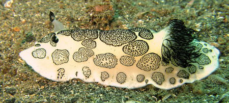
A black-and-white sea slug crawls over an ocean bottom.
This fancy sea slug and its ilk contain anti-cancer compounds in their bodies. Caltech's Brian Stoltz has developed a way to create those compounds in the lab.
Credit: Wikimedia Commons

The natural world, with all its diversity, is a popular place for researchers to go looking for new drugs, including those that fight cancer. 

But there is often a wide gap between finding a plant, sponge, or bacterium that contains a candidate drug, and actually bringing a medicine to the market. Maybe the compound gets flushed out of the human body too quickly to be effective. Or maybe it turns out you have to grind up a metric ton of farmed sea squirts just to get a single gram of the drug. 

For that reason, it usually makes more sense to identify a compound with potential medicinal properties and then make it in the lab, instead of relying on organisms. Often, researchers look to the natural processes that create the compounds for inspiration as they develop synthetic analogs. Though this "biomimetic" method works, it has some limitations. For more than 10 years, Caltech's Brian Stoltz has been looking for a better approach, and now he has found it. 

In December, Stoltz and his research team announced that they had developed a novel synthetic method for creating two compounds that hold the potential to become potent anti-cancer drugs. The compounds, jorumycin and jorunnamycin A, are naturally found only in the bodies of a black-and-white sea slug that lives in the Indian Ocean. 

Both of those compounds are based around a backbone molecule known as bis-THIQ (bis-tetrahydroisoquinoline). In 40 years of research on bis-THIQ compounds, only one has been successfully brought into a clinical setting, Stoltz says. He hopes the production method developed in his lab can change that. 

"We now have a synthesis that's going to let us make whole new compounds," he says. "It's going to enable us to do some really interesting drug-discovery research."

The production method is complex, involving the use of substances called transition metal catalysts, but essentially consists of adding hydrogen atoms to a simpler molecule in a series of steps. The addition of each hydrogen atom causes the molecule to fold further in on itself. When fully folded, the molecule is shaped in a way that makes it prone to bonding to and damaging DNA molecules. Medications that damage DNA might seem counterintuitive, but they are useful for targeting cancer cells. Since cancer cells multiply more quickly than healthy cells, they need to replicate their DNA more often, and are consequently much more sensitive to DNA damage. 

Many compounds can damage DNA, but the trick is developing them into medications that are toxic enough to kill cancer cells, but not so harmful that they kill the healthy cells as well. The ideal medication will stay in the human body long enough to have a therapeutic effect, but not longer than about 24 hours. 

Tailoring a compound to have the traits that make it an effective drug can be done by choosing what Stoltz calls "handles"—the various atoms and groups of atoms that stick off the molecular backbone. By choosing specific handles to put on a compound, researchers can give it the properties they desire. 

This is where Stoltz's production method shines. Some handles interfere with biologically inspired syntheses of bis-THIQ compounds, but almost any handle will work with Stoltz's method, he says.  

"It took us 10 years to get here, but now we can make new analogs in a week," he says. 

Stoltz says Eric Welin, a postdoc on this research team, deserves much of the credit for refining the synthesis into an elegant solution. 

"It was his creativity, drive, and decisiveness that pushed this forward," Stoltz says. "There was a way we could've finished this project that would've been a B-plus solution to the problem, but he pushed for the A-plus version. Eric insisted on developing a method that can produce either "left-handed" or "right-handed" versions of the final compounds at will, rather than the normal 50/50 mixture of both. It is a little like flipping a coin and being able to make it always land on heads." 

He also credited another member of his research team, graduate student Aurapat "Fa" Ngamnithiporn, with doing much of the laboratory work necessary for performing the final synthesis, and continuing to produce novel non-natural analogs.


Brian Stoltz (center) with postdoctoral researcher Eric Welin (left) and graduate student Aurapat "Fa" Ngamnithiporn (right).

Further research will focus on using the synthesis to develop candidate drugs in collaboration with Dennis Slamon, an oncologist at UCLA.  

The paper describing their findings, titled "Concise total syntheses of (—)-Jorunnamycin A and (—)-Jorumycin enabled by asymmetric catalysis," appears in the December 20 issue of Science.

Funding for the research was provided by the National Institutes of Health, the American Cancer Society, the Margaret E. Early Medical Research Trust, the National Science Foundation, and the Teva Pharmaceuticals Marc A. Goshko Memorial Grant Program.

How the Body Fights Cancer and Intruders

$
0
0
New methods for understanding the immune system and how to boost it
News Writer: 
Lorinda Dajose
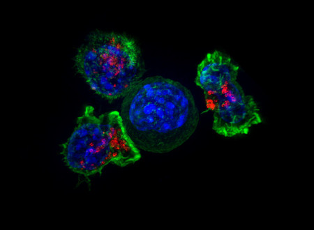
T cells surround a cancer cell
Credit: National Institutes of Health

The human body's immune system is like a vast team of special agents. Certain cells called T cells each individually specialize in recognizing a particular intruder, such as the influenza virus or salmonella. Determining a given T cell's target is a critical step in designing personalized treatments for cancers and developing vaccines. Now, a team of Caltech scientists has developed two new methods for rapidly determining T cell targets.

The work was done in the laboratory of David Baltimore, Robert Andrews Millikan Professor of Biology and president emeritus. Two papers describing the research appear in the January 28 issue of the journal Nature Methods.

A cell that is infected with a pathogen—for example, an influenza virus—will display bits of the invader's genetic material on the cell surface, like waving a red flag to indicate what is going on inside the cell. These "flags," called antigens, are presented on proteins on the cell surface, called MHCs (major histocompatibility complexes). Each T cell is specialized to recognize a different antigen. When a T cell finds a cell displaying its target antigen on an MHC complex, the T cell will bind to it and destroy it.

There are from 1 million to 5 million unique T cells on average in a human targeting countless different pathogens. Though scientists can characterize the function and molecular makeup of a T cell's receptor, it is difficult to determine what target a given receptor specifically recognizes. In fact, fewer than 1,000 antigen–T cell pairs are known.

Now, led by postdoctoral scholars Alok Joglekar and Guideng Li and former postdoctoral scholar Michael Bethune, researchers in the Baltimore laboratory have developed two new methods for determining the targets of T cells.

In the first method, the scientists attached proteins, called signaling domains, onto MHCs. The new complex, called a signaling and antigen-presenting bifunctional receptor, or SABR, is designed to send a signal into the cell to make it glow bright green once it has been bound by a corresponding T cell. A researcher could then take thousands of different antigens, each presented by a SABR, and combine them with a particular T cell. Only the cells presenting the correct antigen should glow green, allowing the researchers to fish out the correct antigen—the T cell's target.

The second method takes advantage of a natural phenomenon called trogocytosis. This occurs when a T cell and its target cell bind together and exchange proteins that are bound to their surfaces. Although researchers have not yet determined why trogocytosis occurs, the Baltimore laboratory decided to use the phenomenon to indicate T-cell targets. To do this, the researchers made a pool of antigen-presenting cells, each displaying a unique antigen, and then exposed them to T cells with a receptor of interest. Only the cells presenting the correct antigen acquired markers from the T cell via trogocytosis. Afterward, the antigen corresponding to the T cell could then be identified by the marker on its surface.

Understanding antigen–T cell pairs is crucial for developing cancer vaccines and also for designing personalized treatments for cancers, as antigens can also be signatures of cancer. No two cancers are the same, and because cancer cells grow so rapidly, they also mutate rapidly. Ideally, then, a scientist could take a sample of a person's tumors, isolate T cells from them, and use one of these methods to discover the antigens that are targeted by the T cells. Once these targets are identified, the T cells can be used to augment the patient's own immune system in various ways to help it better fight the person's cancer.

A paper describing the SABR method is titled "T cell antigen discovery via Signaling and Antigen-presenting Bifunctional Receptors." Postdoctoral scholar Alok Joglekar is the study's first author. In addition to Joglekar and Baltimore, co-authors are Caltech research technicians Michael Leonard and Margaret Swift; former Caltech research technician John Jeppson; postdoctoral scholar Guideng Li; former Caltech undergraduate and current research technician Stephanie Wong (BS '16); former Caltech postdoctoral scholar Songming Peng now of PACT Pharma; Jesse Zaretsky of UCLA; James Heath, a former Caltech professor now at the Institute for Systems Biology in Seattle; Antoni Ribas of UCLA; and former postdoctoral scholar Michael Bethune.

A paper describing the trogocytosis method is titled "T cell antigen discovery via trogocytosis." Guideng Li and Michael Bethune are the study's first authors. In addition to Li, Bethune, and Baltimore, co-authors are Stephanie Wong; Alok Joglekar; Michael Leonard; undergraduate Jessica Wang; former graduate student Jocelyn Kim (PhD '16), now of UCLA; Donghui Cheng of UCLA; Songming Peng; Jesse Zaretsky; Caltech graduate students Yapeng Su and Yicheng Luo; James Heath; Antoni Ribas; and Owen Witte of UCLA. Baltimore, Ribas, Heath, and Witte are also members of the Parker Institute for Cancer Immunotherapy at UCLA and Caltech.

Watson Lecture Preview: Plant Growth—How Stem Cells Make Stems

$
0
0
photo of Elliot Meyerowitz
Elliot Meyerowitz

Plants are the dominant source of our food, clothing, shelter, and many pharmaceutical drugs, yet we know very little about how they live and grow. Elliot Meyerowitz's laboratory studies the collections of stem cells that create stems, leaves, and flowers, focusing on the spiral phyllotaxis pattern, in which successive leaves or flowers appear at angles of roughly 140 degrees. In his February 13 Watson Lecture, Meyerowitz will describe how this pattern forms, answering questions that long have intrigued mathematically inclined biologists and revealing surprising modes of communication between plant stem cells.

Elliot Meyerowitz is the George W. Beadle Professor of Biology and a Howard Hughes Medical Institute Investigator. He is working to understand how plants grow and develop. Much of his research is focused on striving to understand the control of shoot apical meristems—a group of cells that generates a plant's leaves and flowers—with direct relevance to feeding the world and ameliorating the effects of climate change. Meyerowitz received his AB from Columbia University in 1973, and his PhD from Yale University in 1977. He started at Caltech as an assistant professor in 1980, becoming an associate professor in 1985, professor in 1989, and Beadle Professor in 2002. Meyerowitz was chair of the Division of Biology from 2000 to 2010.

The lecture—which will be held at 8 p.m. on Wednesday, February 13, in Beckman Auditorium—is a free event; no tickets or reservations are required.

Named for the late Caltech professor Earnest C. Watson, who founded the series in 1922, the Watson Lectures present Caltech and JPL researchers describing their work to the public. Many past Watson Lectures are available online at Caltech's YouTube site.


This is a Neuron on Nicotine

$
0
0
Nicotine works inside cells to reinforce addiction
News Writer: 
Lorinda Dajose

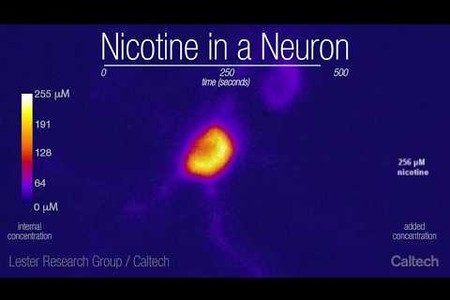
Lester Group - Nicotine in a Neuron

Lester Group - Nicotine in a Neuron
A neuron containing a biosensor developed by the Lester laboratory glows in the presence of varying concentrations of nicotine.
Credit: Caltech/Lester Group

When a person takes a puff on a cigarette, nicotine floods into the brain, latching onto receptors on the surface of neurons and producing feelings of happiness. But nicotine does not simply stay on the surface of cells—the drug actually permeates into neural cells and alters them from the inside out. Now, a team of scientists has developed a protein sensor that glows in the presence of nicotine, allowing the researchers to observe nicotine's movements in cells and reveal more about the nature of nicotine addiction. 

The work was led by Henry Lester, professor of biology at Caltech and previously a visiting scientist at the Janelia Research Campus of the Howard Hughes Medical Institute (HHMI). A paper describing the research appears online on February 4 in the Journal of General Physiology. Lester is also an affiliated faculty member of the Tianqiao and Chrissy Chen Institute for Neuroscience at Caltech.

The endoplasmic reticulum is the equivalent of a cell's factory and warehouse—the place where proteins are synthesized and packaged in order to be shipped to various other locations both inside and outside of the cell. Nicotinic receptors (nAChRs) are among these proteins; after being manufactured in the endoplasmic reticulum, they then travel to the cell's surface. When nicotine molecules enter the body, they travel through the bloodstream and reach brain cells, where they meet the nAChRs on the surface of these cells. This triggers the cells' processes of releasing chemicals of reward and happiness.

What happens once nicotine has moved into the cells, however, has not been well understood. Lester and others previously found that some nAChRs remain in the "warehouse"—the endoplasmic reticulum—where they, too, can bind to nicotine. Hoping to gain insights into nicotine's effects within cells, Lester and his team developed a tool called a biosensor to visualize where the drug collects inside of cells. The biosensor is composed of a special protein that can open and close, like a Venus flytrap, and an inactivated fluorescent protein. The sensor is designed to close around nicotine, and this then activates the fluorescent protein to glow brightly, indicating where the nicotine molecules are located and how many are present. 

Scientists can put the biosensors into particular parts of a cell—in this work, they placed them in the endoplasmic reticulum and on cells' surfaces—and watch them light up as nicotine floods in.

By making movies of cells containing biosensors in a lab dish, the team has discovered that nicotine enters into the endoplasmic reticulum within a few seconds of appearing outside a cell. Furthermore, the nicotine levels are more than enough to affect nAChRs during their assembly and to chaperone additional nAChRs on their journey to the cell surface. As a result, the neurons are more sensitive to the nicotine, which enhances the rewarding feelings after a puff on a tobacco cigarette or an e-cigarette. In other words, the more a person smokes, the more quickly and easily the smoker gets a nicotine buzz. This is part of nicotine addiction.

While the movies now focus on isolated neurons in the lab, the scientists want to determine whether nicotine's intracellular movements are similar in the neurons of live mice. Additionally, they are developing biosensors for other drugs, such as opioids and antidepressants, to observe how these compounds interact inside and outside of cells.

The paper is titled, "Determining the Pharmacokinetics of Nicotinic Drugs in the Endoplasmic Reticulum Using Biosensors." Co-first authors are Amol Shivange, formerly a Caltech postdoctoral scholar and now at Novozymes in Bangalore; Philip Borden of Janelia; and Caltech graduate student Anand Muthusamy. In addition to Lester, other authors are Caltech postdoctoral scholars Aaron Nichols, Kallol Bera, Matthew Mulcahy, and Saidhbhe O'Riordan; Huan Bao and Edwin Chapman of the University of Wisconsin; Ishak Bishara, a former intern in the Lester lab; former Caltech undergraduate Janice Jeon (BS '18); Bruce Cohen, a senior scientist in the Lester lab; Charlene Kim, research technician assistant in the Lester lab; Dennis Dougherty, the George Grant Hoag Professor of Chemistry at Caltech and director of the Beckman Institute; Jonathan Marvin of Janelia; and Loren Looger of Janelia. Funding was provided by the National Institutes of Health, the California Tobacco-Related Disease Research Program, the California Institute for Regenerative Medicine, the Brain & Behavior Research Foundation, HHMI, the Della Martin Foundation, Louis and Janet Fletcher, and Caltech Summer Undergraduate Research Fellowship donors.

Dietary Fiber Helps Clump Material in Your Gut

$
0
0
The results may have implications for microbe growth and drug absorption in our bodies
News Writer: 
Whitney Clavin
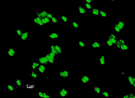
Particles in the small intestinal fluid of mice
Particles in the small intestinal fluid of mice are seen here in 3D. The mice were fed a standard diet.
Credit: Caltech/Ismagilov Lab/A. Preska Steinberg

Food, microbes, and medicine all clump together as they move through our gut. Sticky molecules secreted into our intestines bind the gut particles in the same way that flour holds a ball of dough together. Now a new mouse-based study from Caltech is showing that dietary fiber also plays a role in clumping. This is the first time that researchers have shown that fiber—stringy molecules found in foods like vegetables and whole grains—helps to aggregate gut particles.

"This is the first step in unraveling how the physical properties of fiber impact aggregation in our gut," says Asher Preska Steinberg, a graduate student at Caltech and lead author of a new report in the journal eLIFE. "While sticky molecules in our gut aggregate the particles through chemical binding, the process can be physical with fiber—the fiber molecules cause particles to aggregate by simply sucking out water from in between the particles."

The bundling of particles in our gut may play a role in drug absorption and the regulation of microbe populations, though the details are unclear. Some evidence has shown that particle aggregation can help clear bad bacteria from our gut, while other studies have shown conversely that the clumping can promote colonization, or the overgrowth of bad bacteria. It is also not known if this clumping affects the delivery of drugs or nutrients into our bodies, but some scientists have speculated that it might hinder this process.

"Current dietary guidelines recommend consumption of fiber, but the word 'fiber' is used to describe molecules with a wide range of sizes and other properties," says Rustem Ismagilov, the Ethel Wilson Bowles and Robert Bowles Professor of Chemistry and Chemical Engineering. "Our goal is to understand what each of these different types of fiber are capable of doing in the digestive tract, and the mechanisms responsible for how each type of fiber acts."

In the new study, the researchers fed mice a diet of two different types of fiber: Fibersol-2, a synthetic form of fiber that resembles dietary fiber; and pectin, a fibrous molecule, longer than Fibersol-2, found in apples. They found that pectin, but not Fibersol-2, promoted physical aggregation of particles in the gut. 

"The longer the fiber, the more clumping we saw," says Preska Steinberg. "Our results suggest that aggregation can be controlled by dietary fiber, and may even be tunable by feeding mice fibers of different lengths." 

In the future, the researchers hope to perform similar tests, but using microbes, to find out if the physical forces from fiber can also influence microbial aggregation in the gut. 

The eLIFE study, titled, "High-molecular-weight polymers from dietary fiber drive aggregation of particulates in the murine small intestine," was funded by the Defense Advanced Research Projects Agency, the Army Research Office, the National Science Foundation, and the Jacobs Institute for Molecular Engineering for Medicine. Other authors include Caltech graduate students Thomas Naragon, Justin Rolando, and Said Bogatyrev; and Sujit Datta of Princeton University.


Latest Images